Glaucoma is a group of optic nerve diseases resulting in damage to the optic nerve and ultimately vision loss. In most cases, glaucoma has no symptoms until its late stages. There are many types of glaucoma, some of which are particularly well treated with different types of laser eye surgery.
Glaucoma is the second leading cause of blindness in the U.S. and the leading cause of preventable blindness. According to the American Academy of Ophthalmology, approximately 2.2 million Americans age 40 and older have glaucoma.
Glaucoma, in most cases, can only be detected by regular, routine eye exams. During a routine eye exam, your doctor will do a screening for glaucoma which consists of measuring your eye pressure and examining your optic nerves. If you are determined to be at risk for glaucoma, another testing may be required.
Everyone over age 60 has an increased risk for glaucoma. The risk of having glaucoma is also higher for individuals who have relatives with glaucoma. Heredity, ethnic, and racial background may play a part in determining risk.
There are two broad categories of glaucoma: Open-angle glaucoma and closed-angle glaucoma.
In this type of glaucoma, the drainage angle is open but does not function properly. There are typically no symptoms of this type of glaucoma, and it may only be detected by having a routine eye exam.
Vision loss from open-angle glaucoma tends to happen slowly over the years and generally affects the peripheral vision first. If left untreated, it can result in total loss of vision.
Closed-angle glaucoma is diagnosed when aqueous fluid cannot reach the anterior chamber angle. Closed-angle glaucoma is often painful and sudden, characterized by rapidly progressing visual loss and discomfort.
Sometimes, closed-angle glaucoma can occur due to the scarring of the drainage angle. In these cases, no symptoms may be noted. Symptoms of closed-angle glaucoma include eye pain and redness, blurred vision, headaches, vomiting, and the appearance of halos or rainbows.
Some patients will have a narrow drainage angle that predisposes them to the development of closed-angle glaucoma. Your doctor can easily diagnose this condition during your routine eye exam.
If the drainage angle is very narrow and at high risk for closing, your doctor may recommend a laser procedure called a laser peripheral iridotomy (LPI).
In order to determine if you have glaucoma or if you are at high risk for developing glaucoma, your doctor will perform a series of tests. These tests will need to be repeated at regular intervals to determine if glaucoma is developing or progressing
Your doctor will measure the pressure in your eyes. This is a quick, painless measurement that involves instilling numbing drops in your eyes and measuring your eye pressure using an instrument called a tonometer.
Normal eye pressure readings are typically between 10 and 21, but glaucoma damage can occur at pressures in the normal range.
Since the drainage angle of the eye cannot be viewed directly, your doctor will use a mirrored lens to inspect the drainage angle of your eye.
This will allow your doctor to determine the type of glaucoma you may have and suggest the best treatment options for you.
Inspection of your optic nerve is critical in diagnosing glaucoma because this is where glaucoma damage occurs. This is usually performed with a magnifying lens after dilating drops are placed in your eyes.
Sometimes, your doctor may photograph your optic nerves for monitoring.
This measurement is important to determine the accuracy of your eye pressure measurements. This test is performed after numbing drops are placed in your eyes with a small probe called a pachymeter.
This test helps your doctor find areas of your vision that may be less sensitive due to glaucoma. The test measures your central and side vision. To perform the test, you will be seated with your head positioned in a bowl-shaped device and asked to focus straight ahead.
One eye is tested at a time so an eye patch will be worn on the eye not being tested. You will be asked to press a button when you see flashing lights in your vision while focusing on a target.
Intervention can succeed at many stages in the progress of glaucoma. If you’ve been diagnosed with glaucoma, your doctor will recommend medications, laser, or surgery for the treatment depending on your particular type of glaucoma and lifestyle factors.
Medicated eye drops are the most common way to treat glaucoma. The drops work to lower eye pressure by either improving flow of fluid through the drainage angle or slowing the production of fluid (aqueous humor).
These drops, just like any other prescribed medication, need to be taken regularly. If you have difficulty using drops or if you have unwanted side effects from drops, discuss this with your doctor. There may be other treatment options available for you.
At Mid Ohio Eye, Selective Laser Trabeculoplasty, or SLT is a common way to treat patients with open-angle glaucoma. In SLT, a low-level energy laser targets the drainage angle of the eye and stimulates it to function more efficiently. No holes are created in this process.
This painless procedure takes only minutes to perform and may eliminate or decrease your dependence on eye drops to control your glaucoma. Not every patient will have success with SLT, but if initially successful, this laser can be repeated if the eye pressure rises again.
Laser iridotomy is recommended for patients with closed-angle glaucoma or those with very narrow drainage angles. A laser is used to create a small hole in the iris (colored portion of the eye) in order to allow fluid better access to the drainage angle.
In trabeculectomy, a small flap is made in the sclera (the outer white coating of your eye). A new drainage mechanism is created which bypasses the faulty drainage mechanism.
A filtration bleb, or reservoir, is created under the conjunctiva — the thin, filmy membrane that covers the white part of your eye.
Once created, the bleb looks like a bump or blister on the white part of the eye above the iris, but the upper eyelid usually covers it. The aqueous humor can now drain through the flap made in the sclera and collect in the bleb, where the fluid will be absorbed into blood vessels around the eye.
Eye pressure is effectively controlled in three out of four people who have trabeculectomy. Although regular follow-up visits with your doctor are still necessary, many patients no longer need to use eye drops.
If the new drainage channel closes or too much fluid begins to drain from the eye, additional surgery may be needed.
Aqueous shunt surgery is another type of glaucoma surgery that is usually successful in lowering eye pressure. An aqueous shunt is a small plastic tube or valve connected on one end to a reservoir (a roundish or oval plate). The shunt is an artificial drainage device and is implanted in the eye through a tiny incision.
The shunt redirects aqueous humor to an area beneath the conjunctiva (the thin membrane that covers the inside of your eyelids and the white part of your eye).
The fluid is then absorbed into the blood vessels. When healed, the reservoir is not easily seen unless you look downward and lift your eyelid.
Micro Invasive Glaucoma Surgery, or MIGS, is an important new option in glaucoma management. These types of procedures, allow improvement in the drainage of aqueous fluid in the eye with improved side effect profiles compared to more traditional glaucoma surgeries.
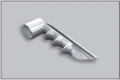
Dr. San Filippo is now performing a type of MIGS procedure, the iStent® Trabecular Micro-Bypass in combination with cataract surgery. The iStent® is the first device of its kind, allowing improvement in the natural outflow of aqueous which lowers eye pressure. The device is the smallest device ever approved by the FDA and it cannot be seen or felt after surgery.
If you have both cataracts and glaucoma and want to know more about iStent®, request an appointment with Dr. San Filippo here.